 Whatsapp
 Facebook
 Twitter
 Instagram
 Linkedin
 Pinterest
 Youtube
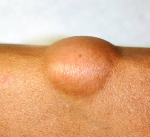
Treatment
Lipoma are benign tumors often, mostly don’t require any treatment other than observation by the patient and their dermatologist, but if a lipoma is painful or continues to grow bigger, it can be removed with a surgical procedure. The only treatment that will completely remove a lipoma is a surgical procedure called excision.
Surgical excision of lipomas often provides a cure.
Procedure
A local anesthetic is injected around the tumor to numb the area to be treated. Large lipomas or those which are deep may require regional anesthesia by injecting numbing medicine into specific nerves. After the anesthesia is given, dermatologist will make an incision in the skin and cut the tumor out, the incisions are configured like a fusiform excision following the skin tension lines and are smaller than the underlying lipoma. Gradual dissection is then performed beneath the subcutaneous fat to the lipoma and tissue cutting is performed using a no.15 scalpel or scissors around the lipoma. Once a part of lipoma has been dissected from the surrounding tissue, clamps can be attached to the lipoma to provide traction for removal of the remaining growth. Once lipoma is freed, it is taken out as a whole.
Hemostasis is achieved following the removal of the lipoma using suture ligation. The dead space is closed beneath the skin using buried, interrupted sutures. The skin is then closed with interrupted sutures. After excision, a pressure dressing is placed to reduce the incidence of hematoma formation.
Post Procedure
Patient should be able to go home soon after the procedure if it is a small or superficial lipoma. The routine wound care instructions usually given along with oral antibiotics, and the wound is checked after two to seven days. The sutures are usually removed after 7 to 14 days. Specimens should be sent for histopathological analysis to rule out any malignancy.